- Services & Solutions
- Technologies
- Discovery
- Reagent Materials Generation
- Monoclonal Antibody Discovery
- Bispecific & Multispecific Antibody Generation
- Lead Optimization
- In Vitro Lead Characterization
- In Vivo Lead Characterization
- Other Molecular Modalities
- IND Filing Support
- Protein Sciences
- Antibody Production
- Protein Production
- Protein Characterization
- Protein Sciences
- Mammalian
- Microbial
- mRNA
- Viral Vaccines
- WuXi XDC – Bioconjugation
- Testing
- News & Resources
WuXi Biologics
Offering End-to-End Solutions
- Services & Solutions
 News & Resources
 - News & ResourcesNews & Resources


>
In Vivo Lead Characterization
The WuXi Biologics discovery services team has years of experience and provide well-trained experts in the wide variety of platforms and technologies offered for in vivo characterization of preclinical candidates (PCCs). We have supported 800+ projects and have successfully progressed many products into IND filing stage, clinical trials and on to commercialization.
Our senior scientists and experts, many trained in the U.S. and Europe, provide the full-spectrum of in vivo characterization services including in vivo efficacy evaluation of therapeutic monoclonal, bispecific and multispecific antibodies, ADCs, CAR-T products, and other large molecules for the treatment of tumor, autoimmune conditions, inflammatory indications, diabetes and other diseases. We also provide high-quality preclinical PK, safety, exploratory toxicology, biomarker and ex vivo studies to support pharmaceutical companies worldwide.
In Vivo Efficacy Studies
Characterization and evaluation of biotherapeutic efficacy is critical for lead candidate selection. We have established a comprehensive database on the effectiveness and utility of cell lines, targets, and models based on client need, and indications over the past 10+ years. We have access to a wide range of models and provide professional consultation and expertise to select the best model for your program based on your specific project requirements. Our high-quality screening platforms include a variety of models for efficacy assessments in the following disease areas:
- Oncology
- Other (non-cancerous) tumor generating conditions
- Autoimmune disease and inflammation
- Metabolic disease
- Graft-versus-host disease
- Rare diseases (to be determined based on request)
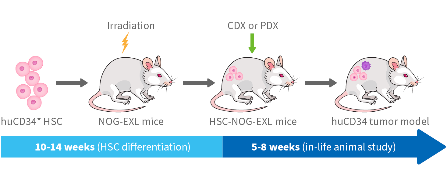
Tumor Models
WuXi Biologics provides an extensive variety of tumor models for efficacy characterization of preclinical candidates (PCCs) that include:
Syngeneic models use syngeneic tumor cell lines and the inbred strain as a recipient.
Human cell line-derived xenograft (CDX) models are an efficient and cost-effective means to obtain efficacy data and information especially for antibodies that do not display murine cross reactivity. These models allow lead candidates to easily advance into IND filing and clinical development. WuXi Biologics’ team has designed and validated an extensive list of CDX models.
KI models are unique tools to investigate antibody efficacy on the human immune system or to interpret the antibody responses to tumor-associated antigens (TAA), to which the antibody drug has no cross-reactivity. We offer a wide variety of single, double or triple KI models for antibody evaluation. Below is a list of KI models we have evaluated for use in biotherapeutic screening.
Single target:
- h-PD1, hPD-L1, hSIRPA, hCD47, hBTLA, hCD28, hCTLA4, hGITR, hOX40, hTIGIT, hTIM3, hLAG3, h4-1BB, hCD27, hCD3E, hCD94, hCD73, h4-1BB-L, hB7-H3, hCD38, hNKG2A, hSIGLEC10, hLRRC33, hCD226.
- HIL7R, B-hCXCL13, B-hRSPO1, B-hIL21R, hIL13, hCD40, hOX40, hTNFR2, hCD28, CD20, CTLA4, hIL17A, hTNFA, HIL6, hIL33, hIL1B, hIL1B, hIL4, hIL4RA, etc.
Double or triple target knock-in
-
h4-1BB/h4-1BB-L, hCTLA4/h4-1BB, hCTLA4/hLAG3, hCTLA4/hOX40, hCTLA4/hTIM3, hOX40/h4-1BB, hPD-1/h4-1BB, hPD-1/hBTLA, hPD-1/hCD40, hPD-1/hCTLA4, hPD-1/hGITR, hPD-1/hLAG3, hPD-1/hOX40, hPD-1/hPD-L1, hCD47&hSIRPa, hPD-1/hTIGI, hPD-1/hTIM3, hPD-L1/h4-1BB, hPD-L1/hCD40, hPD-L1/hCTLA4, hPD-L1/hLAG3, hPD-L1/hOX40, hPD-L1/hTIM3, hPD-L1/hTIGIT, hPD-L1/hSIRPa/hCD47, hPD-1/hPD-L1/h4-1BB, hPD-1/hPD-L1/hCTLA4, hPD-1/hPD-L1/hTIGIT, hPD-1/hPD-L1/hTIM3, hPD1/hLAG3/hTIM3, hPD-1/hPD-L1/hTNFR2, hPD-1/hPD-L1/hCD40, hLTA/hTNFA/hLTB etc.
-
hIL17A/hIL17F, hTNFA/hIL17A, B-hIL4/hIL4RA, B-hIL12A/hIL12B, hIL6/hIL6R, hIL23A/hIL12B etc.
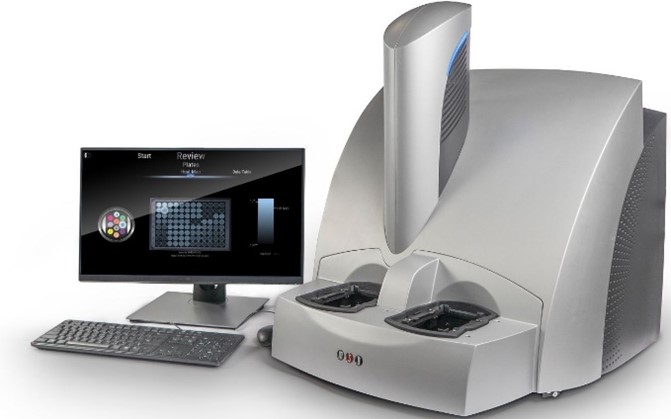
WuXi Biologics utilizes the IVIS Spectrum in vivo 2-D and 3-D imaging system as an advanced tool in the characterization of preclinical oncology candidates including CAR-T therapies. Using this platform, we also specialize in providing screening services for the evaluation of systemic tumors and, metastasis.
IVIS Spectrum Capabilities
- High sensitivity 2D in vivo optical imaging of bioluminescence and fluorescence tagged molecules
- High-sensitivity 3D in vivo optical tomography for bioluminescence & fluorescence imaging
- High-throughput (5 mice simultaneously) imaging with 23 cm field of view
- High-resolution (to 20 microns) imaging with 3.9 cm field of view
- High-efficiency wavelength filters spanning 430-850 nm
- Ideal for distinguishing multiple bioluminescent and fluorescent reporters
IVIS Spectrum imaging application – Bioluminescent in vivo imaging
The IVIS Spectrum system is unique in the evaluation of orthotopic, systemic and metastasis models. The bioluminescent tumor models provide a clinically relevant, organ-specific tumor microenvironment, and enhance accuracy and relevancy over subcutaneous models. The models, used in this platform mimic primary tumor lesions, have better interactions among tumor, stroma and immune components, and obtain results more relevant to tumor progression and spontaneous metastasis.
Therefore, bioluminescent models provide an ideal setting to track disease progression in real time and investigate drug anti-tumor efficacy as well as mechanisms.
The WuXi Biologics team have years of experience and have established a variety of validated models in the IVIS Spectrum system.
We provide a number of disease models to characterize monoclonal, bispecific and multi-specific antibodies and protein drugs targeting autoimmune and metabolic diseases. Many of the models available are as follows:
Autoimmune Encephalomyelitis (EAE)
- MOG35-55-induced EAE in C57BL/6
- MBP-induced EAE in Lewis rats
- PLP131-135-induced EAE in SJL/J
Rheumatoid Arthritis (RA)
- Collagen-induced arthritis (CIA) in DBA/1
- Collagen-induced arthritis (CIA) in Lewis rats
- Adjuvant-induced arthritis (AIA) in Lewis rats
Asthma model
- OVA-induced asthma in BALB/c
- HDM-induced asthma in C57BL/6
Systemic Lupus Erythematosus (SLE)
- Pristane-induced lupus in BALB/c
- MRL/lpr mice lupus model
Intestinal Bowel Disease Model (IBD)
- DSS-induced colitis in C57BL/6
Air pouch Model
- Carrageenan-induced air pouch in BALB/c
- Carrageenan-induced air pouch in Wistar rats
- rhIL-13-induced air pouch in BALB/c
Atopic Dermatitis (AD) and Psoriasis model
- DNCB-induced dermatitis in C57BL/6
- DNCB-induced dermatitis in Brown Norway rats
- Oxazolone-induced atopic dermatitis in BALB/c
- MC903-induced atopic dermatitis in C57BL/6
- Imiquimod-induced psoriasis in BALB/c
- IL-23-induced ear epidermal hyperplasia in C57BL/6
- KLH-induced DTH in BALB/c
- Oxazolone-induced DTH in BALB/c
- hIL-31-induced pruritus in hIL31/hIL31RA/hOSMR
Fibrosis and liver injury model
- Bleomycin-induced pulmonary fibrosis in C57BL/6
- Folic acid-induced renal fibrosis in C57BL/6
- APAP-induced liver injury in C57BL/6
T cell dependent antibody response (TDAR) model
- KLH-induced TDAR in C57BL/6
Eosinophilic Gastroenteritis (EG)
- OVA-induced Eosinophilic Gastroenteritis in C57BL/6
PK/PD and Exploratory Toxicology Platforms
Pharmacokinetics (PK)
Pharmacokinetics (PK) is the study of drug absorption, distribution, localization, metabolism, biotransformation and excretion. The primary goal for antibody PK studies is to identify drug candidates that demonstrate ideal PK characteristics that potentially enhance efficacy with decreased toxicity. The data generated for lead candidates provide key reference for the IND filing and clinical studies. With extensive experience and capabilities, we have established a substantial list of PK assays for protein drug candidates.
Capabilities
- A variety of ELISA methodologies validated for consistency and accuracy
- ELISA via coating antigens of Fc, Fab1, ADC-payload, TCR, cytokine, VHH and albumin with detection antibody of Fc, VHH or Fab2
- MSD is available for micro-scale sampling with high accuracy and sensitivity
- LC-MS for ADC analysis
- Data interpretation using industry standard WinNonlin software
Animal Species
- Mouse
- Rat
- Rabbit
- Monkey
- Canine (Under development)
- Swine (Under development)
PK parameters
|
Parameter |
Definition |
Unit |
|
Cmax |
Maximum observed concentration |
μg/mL |
|
Tmax |
Time of maximum observed concentration |
h |
|
AUClast |
Area under the concentration-time curve |
μg/mL*h |
|
Vd |
Apparent volume of distribution |
mL/kg |
|
Cl |
Volume of plasma cleared of the drug per unit time |
mL/h/kg |
|
T1/2 |
Half life |
h |
|
MRT |
Mean residence time |
h |
Pharmacodynamics (PD) and Exploratory Toxicology
Drug safety is an important step in therapeutic antibody development. We have experts with years of experience in this field and provide high quality solutions and service for antibody safety assessment. Early stage PD and toxicological tests combined with toxicokinetic studies give more information for non-clinical studies, phase I dose selection, toxic reactions and severity analysis, etc. In some cases, short-term subacute toxicity tests (1-3 months) convey safety issues, and help determine whether the innovative candidate can progress towards IND filing and early clinical trials.
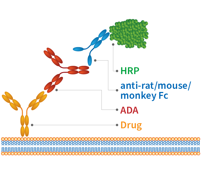
ADA Analysis
In preclinical models and clinical trials, antibodies often elicit Anti-Drug Antibodies (ADA), which can then interfere with antibody efficacy evaluation. Thus, ADA detection and evaluation facilitates understanding of potential immune responses to biologic drugs as well as potential risks. We provide solutions to determine ADA production at discovery and preclinical stage and our services include:
- Generation of detection antibodies and solutions
- Detection of ADA against conjugated drugs
- Detection of anti-drug neutralizing antibody
- Production and detection of anti-idiotype antibody
Ex Vivo Platforms
WuXi Biologics provides a series of ex vivo platforms, assays and methodologies for the evaluation and characterization of drug candidates. Examples of our vast capabilities include cytokine assays, receptor occupancy (RO) assays, histopathology, FACS, hematology and clinical chemistry.
- Histology
- FACS
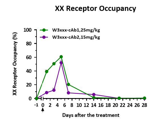
- Receptor Occupancy (RO) Assay
- Cytokine Assays
- Clinical Chemistry (CHEM)
- Hematology (HEM)
- Coagulation Analysis for Mematological Diseases
- MSD-ECL Detection System
We provide comprehensive, high-quality pathology services utilizing advanced instruments and equipment. The labs are equipped with paraffin embed machines, paraffin microtomes, cryostat sectioning, blocks sample storage, slides sample storage, -80° C degree sample storage, fume cupboard, and fluorescent and light microscopes with digital photography and quantitative analysis.
Histology laboratory capabilities
Tissue bank in paraffin-embedded block
- Murine samples
- Monkey samples
- Human samples
Slides type
- Formalin-fixed paraffin embedded (FFPE)
- Optimal cutting temperature compound embedded (OCT)
- Cell smear sample
Staining type
- Immunohistochemistry (IHC/IHC double)
- Immunofluorescence (IF/IF double)
- Enzyme histochemistry (EHC)
- Tissue bank for TCR (Tissue Cross-reactivity)
- Antibody verification for IVD reagent
- Tunnel
- Diff quick
- Masson
- Toluidine blue
- Hematoxylin and eosin(H&E)
- Periodic Acid-Schiff (PAS)
- Oil red
- Luxol fast blue (LFB)
- Safranin O
- Sirius red
Image analyses with HALO system
- Area Quantification
- Multiplex IHC cell counting
At WuXi Biologics we utilize multiple fluorescence activated cell sorting (FACS) systems to perform a wide range of ex vivo studies to assist in lead candidate screening.
Equipment
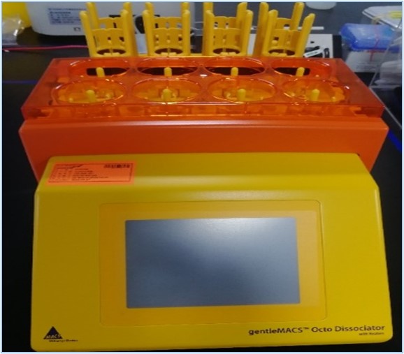
- GentleMACS Octo Dissociator: Single cell suspension preparation with efficiency.
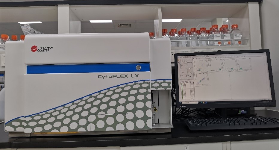
- Beckman Coultor cytoFLEX LX: Multiple colored, 19 channel, high throughput.
Capabilities of FACS
- hPBMC reconstitution detection
- Cell surface receptor occupancy
- Cell killing, cell apoptosis
- TILs analysis
- TAM analysis
- Cytokine CBA assay
- Immuno-phenotyping
Receptor Occupancy (RO) on the corresponding target is quantified with fluorescent- labeled antibody to detect the percentage of unoccupied receptors from in vivo samples or cell lines.
Cytokines are biomarkers for efficacy and potential side effects, such as Cytokine Release Syndrome (CRS). CRS is a systemic inflammatory response caused by disease complications, infections or antibody administration or other adverse effects of biological therapies. Therefore, studies to evaluate CRS resulting from drug candidates is a critical part of PCC screening. We routinely run assays for Th1, Th2, Th17 and CRS cytokine detections with high-speed and accuracy.
CRS detection assays available
- Customized cytokine detection
- MSD multiplex Assay
- FACS CBA multiplex Assay
- ELISA Assay
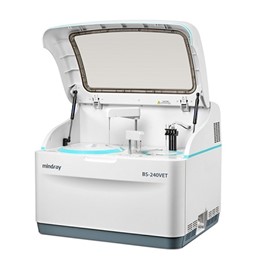
The results from Clinical Chemistry (CHEM) studies are important indicators reflecting the metabolism or content of carbohydrates, lipids, and proteins in urine/kidney, liver, gallbladder, cardiovascular, pancreatic and gastrointestinal tract samples. The indicators provide us data on the toxicological and biological significance of the PCCs. We have accumulated extensive experience in CHEM testing, and provided comprehensive services that meet our customer’s needs.
Chemistry testing with BS-240VET Analyzer
- Liver function: ALT, AST, ALP, T-Bil-D II, TP II, ALB, γ-GT, etc.
- Kidney function: UREA, CREA, MALB, etc.
- Myocardial enzyme spectrum: CK
- Glucose metabolism: Glu, β-HB, etc.
- Fat: TC, TG, etc.
- Pancreas: α-AMY
- Complement proteins: C3, C4 (under development)
- Rheumatism: CRP, ASOII (under development)
- Anemia: Fe, FER (under development)
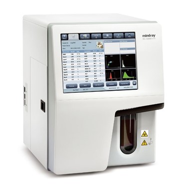
Hematology (HEM) tests are used as an auxiliary means for disease diagnosis and drug safety evaluation. The assays mainly include red blood cell count, white blood cell count, platelet count and hemoglobin content. By analysis of blood test data, we provide preliminary safety evaluation and offer conclusions on the use of the therapeutics being evaluated.
Detection technology
- Auto Hematology Analyzer (BC-5000Vet)
Types of HEM testing
- White blood cell counts: WBC, Neutrophils, Lymphocytes, Monocytes, Eosinophils Basophils and NK cells
- Red blood cells: RBC, HGB, HCT, MCV, MCH, MCHC, RDW-CV, RDW-SD
- Platelets: PLT, MPV, PDW, PCT
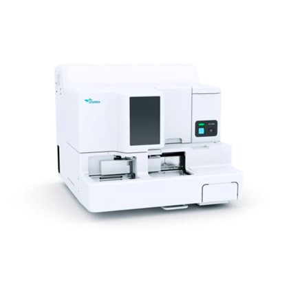
Coagulation Analysis for Mematological Diseases
Coagulation function tests evaluate clot formation after activating the coagulation cascade. The coagulation function system used at WuXi Biologics offers pre-analytic sample checks and four methods simultaneously on a single platform including coagulation end-point, chromogenic kinetic analysis, turbidity immunoassay and automated platelet aggregation.
Detection technology
- Automatic hemostasis analyzer Sysmex CS-2400 analyzer
Types of testing
- Operation: automatic
- Analysis mode: chromogenic, immunologic
- Sample type: whole blood
- Coagulation: PT, APTT, Fbg, TT, PS, PC, LA1, LA2
- Exogenous coagulation factors: II, V, VII, X
- Endogenous coagulation factors: VIII, IX, XI, XII
- Chromogenic kinetic analysis: AT III, αTPI, Plg, PC, FXIII, Hep, C1
- Turbidity immunoassay: D dimer, FDP, ADP, Col and Epi
Electrochemiluminescence immunoassay (ECLIA) is a specific chemiluminescent reaction initiated by electrochemistry on the electrode surface.
The MSD-ECL platform uses an electrochemiluminescent-labeled antibody conjugate for detection purposes. The label usually uses a SULFO-TAG marker. After the electrode surfaces of MULTI-ARRAY and MULTI-SPORT microplates are electrified, the electrochemical action excites the SULFO-TAG marker to emit a strong light. Utilizing the MSD S600MM system with Methodical Mind Software, we significantly facilitate the bioanalyses of our PK, ADA, and Cytokine platforms.
Advantages
- High Sensitivity: low limits of detection and broad dynamic ranges
- High Stability: consistent results from single-factor and multi-factor detection
- Small sample volume: ≤50ul
- Broad Dynamic Range: 4-5 log
- Multiplexed: high throughput, cost-effective for two or more analytes
Key Applications
- Biomarker detection
- Biological drug metabolism
- Immunogenicity
- High-throughput antibody affinity assay
- Signal pathway phosphorylation detection
- High throughput with 384 well plates
Immunization Platform
Hybridoma technology is a classic technology in monoclonal antibody development. We have state-of-art facilities and experts to provide timely and high quality services to our clients.
Advantages:
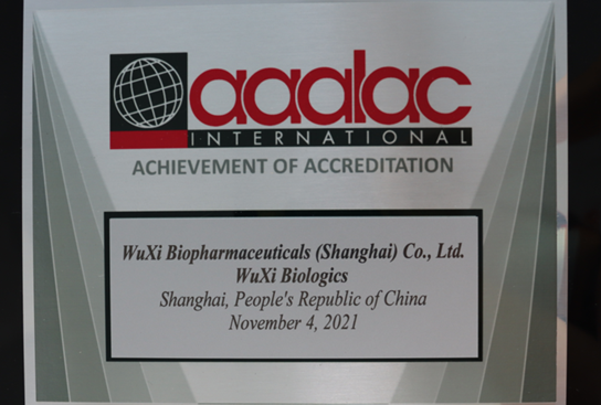
- Certified facilities (e.g., AALAC)
- Highly effective and high quality immunization strategies and protocols (utilizing our own internally developed intellectual property)
- Immunization strategies for DNA, polypeptide / protein, cells, liposome, nanoparticle or combinations of these various antigens
- Ligand’s OmniAb® and Alloy Therapeutics’ ATX-Gx™ platforms to provide humanized monoclonal antibody development services.
DNA-encoded antibody delivery
Monoclonal antibodies have been widely used in the treatment for tumor, inflammatory and autoimmune disorders. However, high costs, and dose frequency limit patients’ accessibility to these therapeutic modalities. DNA delivery presents a labor- and cost-effective alternative to antibody therapeutics, with less frequent delivery and simpler formulation. Nanoparticle delivery systems overcome the biological barriers in vivo, and prolong circulation and improve antibody expression and sustainability. Thus, DNA delivery provides a viable, long-term treatment option. At WuXi Biologics, we have an experienced team to perform in vivo and ex vivo studies to identify lead candidate in this new area for biologics.

Facilities
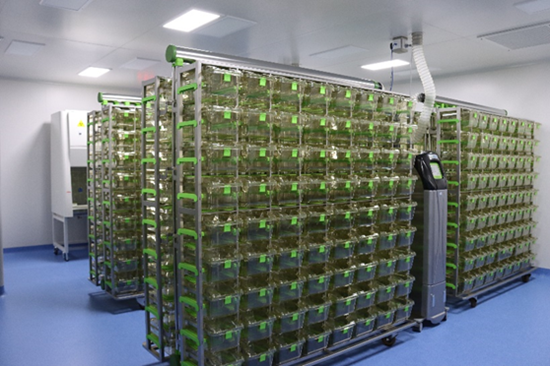
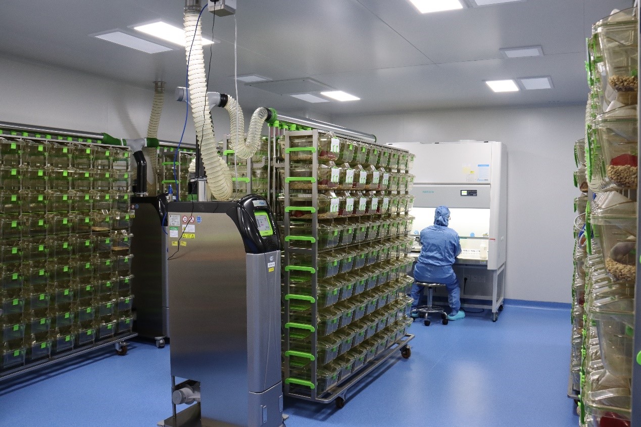
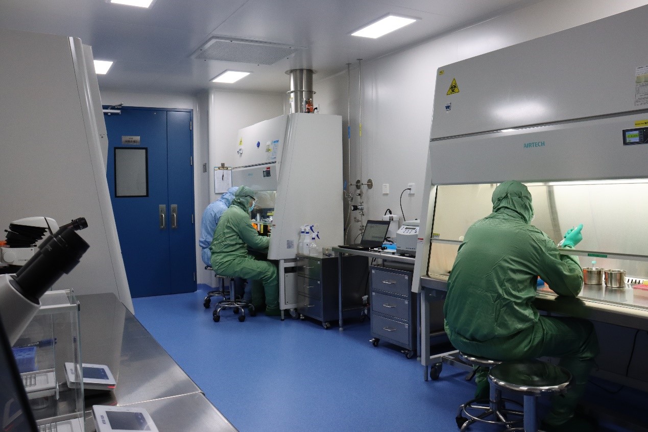
We have a state-of-art SPF facility located in Shanghai with AAALAC accreditation and a License of Laboratory Animal Use obtained in 2021. The facility is equipped with Tecniplast Green Line IVC systems, pit-type pulsating vacuum sterilizer, AⅡ/ BⅡ biosafety cabinet, advanced irradiator, and PE IVIS Spectrum instruments.